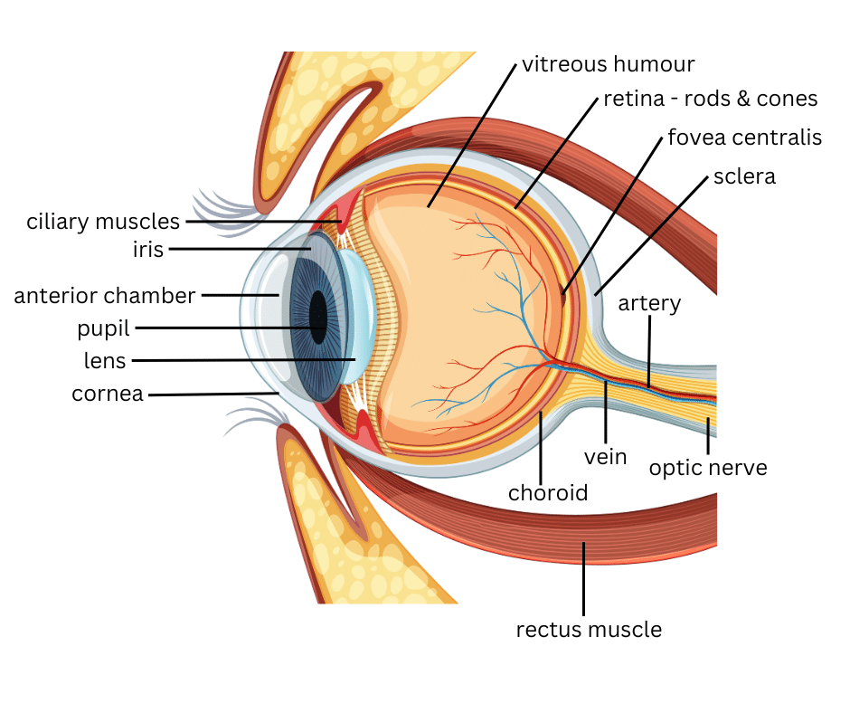
The anatomy of the human eye
The human eye possesses astonishing capabilities. Despite its diminutive size, it undeniably confers upon us the most vital of our five senses – the gift of sight.
Through the intricate interplay of its components, such as the iris and cornea, the eye ingeniously guides the entry of light into its depths, ensuring an optimal amount reaches the lens.
Much akin to the lens of a camera, which orchestrates the creation of a film, the eye’s lens bends incoming light, expertly projecting it onto the retina. This remarkable structure, composed of countless specialised cells known as rods and cones, seamlessly converts the visual stimuli into electrical energy. Subsequently, this newfound energy embarks on a journey, traveling to the optic disk on the retina, where it is then transmitted via electrical impulses along the optic nerve, destined for intricate processing within the confines of the brain.
The eyeball encompasses a trinity of layers to maintain its intricate structure:
- The outermost layer, comprised of the cornea and sclera, acts as a protective shield.
- Nestled in the middle layer, we find the vital bloodstream supply and the captivating iris and pupil.
- Deep within lies the innermost layer, known as the retina, responsible for capturing the essence of vision.
Not only does the eyeball comprise these layers, but it also houses a triad of fluid-filled sanctuaries:
- The anterior chamber, harmoniously situated between the cornea and iris.
- The posterior chamber, a tranquil space nestled between the iris and lens.
- The vitreous chamber, a serene sanctuary between the lens and retina.
The anterior and posterior chambers are bestowed with the presence of aqueous humour, a nourishing aqueous fluid that delicately sustains the inner sanctum of the eye, ensuring its buoyancy. As for the vitreous chamber, it cradles a thicker essence called vitreous humour, an unassuming yet transparent gel, predominantly composed of water, preserving the eye’s delicate equilibrium.

What makes up the human eye?
Choroid
The eye is composed of various components, including the choroid. Situated between the retina and the sclera, the choroid plays a crucial role in maintaining optimal vision. It is a porous layer, resembling a sponge, which serves as a protective barrier.
Furthermore, the choroid contains a specialised pigment that effectively absorbs any surplus light, thereby preventing the occurrence of blurry vision. This element acts as a natural filter, ensuring that the incoming light is appropriately received and processed by the eye.
Additionally, the choroid is densely populated with blood vessels, which serve the essential function of nourishing the outer layers of the retina. Through this intricate network, vital nutrients and oxygen are transported, contributing to the overall health and functionality of the eye. Thus, the choroid, with its unique characteristics and significant responsibilities, remains a vital component in the complex structure of the eye.
Ciliary Muscles
The ciliary muscles, situated at the back of your iris and in close proximity to the crystalline lens, play a vital role within your eye. They serve a dual purpose, first by producing the aqueous fluid that fills the anterior portion of your eye.
Additionally, these muscles possess the remarkable ability to adjust the eye’s focus, enabling it to adapt and see clearly at varying distances.
Cone Cells
Cone cells, which reside in the retinal layer of the eye, comprise the secondary category of light-sensitive cells. Within the human retina, an impressive count of approximately six to seven million cones can be found.
These cells showcase their optimal functionality in well-lit environments, playing a crucial role in facilitating acute vision and ensuring the reception of precise, sharp images.
Scientists hypothesise the existence of three distinct types of cones, each exhibiting sensitivity to the wavelength of a primary colour, namely red, yellow, or blue. Consequently, the human eye perceives all other colours as intricate combinations of these fundamental hues.
Cornea
The cornea, a translucent circular structure positioned at the forefront of the eyeball, plays a crucial role in the visual system. Its main purpose lies in bending and directing incoming light onto the lens, ultimately enabling the lens to properly focus it onto the retina.
Remarkably, the cornea lacks any blood vessels, rendering it highly susceptible to pain sensitivity. Serving as a transparent portal, the cornea encompasses a substantial one-sixth portion of the outermost layer of the eye, precisely designed to concentrate and transmit light, allowing for optimal visual perception.
Conjunctiva
The conjunctiva, a delicate and transparent membrane, envelops the sclera—a prominent component of the eye that is visually represented by its distinctive white hue.
This specialised covering, akin to the protective integument of the eye socket, serves the essential purpose of safeguarding and moistening the eyeball, ensuring its smooth functionality.
Crystalline Lens
The crystalline lens, a remarkable transparent structure nestled within the eye and positioned just behind the iris, assumes the pivotal role of meticulously directing the path of incoming light onto the delicate surface of the retina.
Fovea Centralis
The fovea, located within the macula, presents itself as a modest concavity and harbours the highest density of cone cells. As the eye fixates on an object, the brain meticulously records the image that falls precisely on the fovea, ensuring unparalleled accuracy in perception.
Iris
The iris, a crucial component of your eye, plays a pivotal role in controlling the influx of light. Positioned delicately between the cornea and the crystalline lens, this vibrant structure acts as a barrier, effectively dividing the front and back chambers of the eye.
In its remarkable function, the iris skilfully manages the quantity of light that permeates your eye, ensuring optimal visual conditions.
Lens
The lens, found just behind the pupil, is a clear structure surrounded by a delicate, see-through casing. Its primary function is to bend and concentrate incoming light, directing it precisely onto the retina.
However, when the lens becomes hazy, it can lead to a condition known as cataract. In circumstances where a cataract impairs vision significantly, a surgical intervention is performed to substitute the cloudy lens with a synthetic lens made of plastic.
Macula
The macula, a region of vivid yellow hue, gracefully envelops the fovea located at the posterior of the eye’s retina. Positioned with great precision near the heart of this ocular structure, the macula is a diminutive yet exquisitely perceptive segment responsible for the intricate nuances of our central vision.
As the epicentre of the macula, the fovea enables us to luxuriate in the richness of detail, empowering us to execute essential activities that necessitate focused sight, such as devouring words on a page or manoeuvring a vehicle with confidence.
Optic Disk
The optic disc, observable during eye examinations, is a distinct section of the optic nerve that can also be detected on the retina. It serves as a marker for the initial point of the optic nerve, through which signals generated by cone and rod cells exit the eye via nerve fibres, ultimately reaching the optic centre of the brain. This region is commonly referred to as the “blind spot.”
Optic Nerve
The optic nerve emerges from the eye at the optic disc, diligently conveying all visual data to the brain. This vital nerve, nestled at the posterior of the eye, plays a crucial role in transmitting the intricate images perceived by the retina to the brain. The optic disk, discernible on the retina’s outer layer, marks the anterior surface of the optic nerve.
Countless nerve fibres intricately interweave within the retina, amalgamating to form the optic nerve. As light penetrates the retina, it metamorphoses into electrical signals, which traverse these fibres, embarking upon a transformative journey through the optic nerve, ultimately reaching the brain.
Pupil
The iris of the eye encompasses a circular aperture known as the pupil, acting as a gateway for light to reach the lens. Controlling the expansion and contraction of the pupil, the iris governs the phenomenon of dilation and constriction.
The pupil, residing within the iris, serves as a sombre focal point. Its primary purpose lies in the meticulous management of light intake into the eye. By means of an intricate mechanism, the pupil autonomously adjusts its dimension to ensure a harmonious equilibrium of luminosity permeating the ocular realm.
Retina
The retina, an intricate layer that coats the innermost part of the eye, holds the remarkable ability to detect and respond to light. Comprising specialised cells called rods and cones, this light-sensitive marvel is instrumental in our visual perception.
Rods, numbering an impressive 125 million in the human eye, play a crucial role in facilitating vision in low-light conditions. Conversely, cones, numbering between six and seven million, are most proficient in capturing vibrant details under bright illumination and enabling colour differentiation.
In a manner akin to the film within a camera, the retina astoundingly translates light into electrical impulses, which then travel along the optic nerve to the brain, culminating in our perception of the world around us.
Sclera
The sclera, commonly known as the eye’s white region, constitutes approximately eighty-three percent of the eye’s outer layer. Its primary role revolves around safeguarding the delicate structures of the eye, and it also serves as the anchoring point for the extraocular muscles that facilitate eye movement.
Vitreous Humour
The vitreous humour is a transparent gel-like substance occupying the central region of the eye, providing structural support. It primarily consists of water and encompasses approximately two-thirds of the eye’s overall volume.
This vital component aids in preserving the eye’s spherical form and is intertwined with the retina at multiple locations, such as the macula and the optic nerve.
Comprehensive Solutions for Optical Ailments
Medical Eye Clinic in Exeter, Devon presents a pioneering concept, bringing together a team of esteemed ophthalmologists and optometrists dedicated to enhancing eye care in the region.
A cutting-edge surgical facility, integrated with one of the most reputable optician chains in the South West, Bill Opticians, sets the stage for the exceptional standards of eye care delivered by The Medical Eye Clinic. Our commitment remains unwavering in providing unparalleled, professional services. Whether you seek an elevated level of eye examination, wish to explore the realm of contact lenses, or require new spectacles, we are poised to address your needs comprehensively. Even if you harbour concerns regarding any optical ailments, rest assured, our expertise shall guide you towards resolution.
Through a diverse range of eye care professionals, we strive to curate an unparalleled experience, placing your satisfaction at the heart of our practice. By harnessing state-of-the-art technology and diagnostic instruments, we ensure that our eye examinations and specialised procedures attain utmost thoroughness and precision.